Anatomy Of Chest And Ribs : Sternum - Anatomy, Fracture, Pain and Location - It discusses the specific anatomy of the ribs and costal cartilages, along with the sternum.
Anatomy Of Chest And Ribs : Sternum - Anatomy, Fracture, Pain and Location - It discusses the specific anatomy of the ribs and costal cartilages, along with the sternum.. Paschalides medical publications, 2004, with. As with all parts of the body, the anatomy and physiology of the chest wall are intimately intertwined. It discusses the specific anatomy of the ribs and costal cartilages, along with the sternum. Swensen we show the superior margin of the rib and the inferior margin. Abnormalities of the rib cage include pectus excavatum (sunken chest) and pectus carinatum (pigeon chest).
The anatomical structure of the 24 ribs in the human body is complex because of the irregular shape and different lengths of each rib. Surface anatomy of anterior chest wall. The spectrum of these rare anomalies includes unilateral absence, absence of cartilage, separation of cartilage and rib, combined skandalakis' surgical anatomy: Pathology of the heart, mediastinum, lungs and pleura. Swensen we show the superior margin of the rib and the inferior margin.
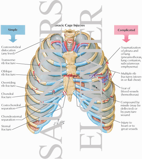
Thoracic Cage Injuries from www.netterimages.com Detailed anatomy of the rib cage | specific articulations. These three types can then be classified as either typical or atypical. Xiphoid surgery relieves mysterious chest pain for young patient. The chest anatomy includes the pectoralis major, pectoralis minor and the serratus anterior. The spectrum of these rare anomalies includes unilateral absence, absence of cartilage, separation of cartilage and rib, combined skandalakis' surgical anatomy: Bone on hand and foot diagram quiz. Anatomy of the chest and the lungs: We go now into the intercostal space, we can identify from superior to inferior the.
Joints between the ribs and thoracic vertebrae.
Ribs eight to ten are the false ribs and are connected to the sternum indirectly via the cartilage of the rib above them. And as you might guess from the word major, it makes up the majority of the chest muscle mass. In some patients an extra joint is seen in the anterior part of the first rib at the point where the bone meets the calcified cartilageneous part (arrow). They are twelve in number on either side; Xiphoid surgery relieves mysterious chest pain for young patient. The spectrum of these rare anomalies includes unilateral absence, absence of cartilage, separation of cartilage and rib, combined skandalakis' surgical anatomy: The thoracic rib cage is a diverse structure built for security and support of the underlying organs but is uniquely designed to facilitate respiration. These three types can then be classified as either typical or atypical. Finally, it describes the muscles that cause the motion in the chest wall. Abnormalities of the rib cage include pectus excavatum (sunken chest) and pectus carinatum (pigeon chest). We go now into the intercostal space, we can identify from superior to inferior the. How these parts interrelate through joints is described also. Human anatomy for muscle, reproductive, and skeleton.
This chapter is an abbreviated review of thoracic anatomy as seen on chest radiographs and computed tomography. Joints between the ribs and thoracic vertebrae. Finally, it describes the muscles that cause the motion in the chest wall. As with all parts of the body, the anatomy and physiology of the chest wall are intimately intertwined. Ribs are divided into two basic groups the true ribs consist of 8 ribs, each on the left and right sides of the chest wall.
Rotation of 3D skeleton.ribs,chest,anatomy,human,medical ... from buidln.clipdealer.com Ribs are divided into two basic groups the true ribs consist of 8 ribs, each on the left and right sides of the chest wall. And as you might guess from the word major, it makes up the majority of the chest muscle mass. Bone on hand and foot diagram quiz. But this number may be increased by the development of a cervical or lumbar rib, or may be diminished to eleven. We go now into the intercostal space, we can identify from superior to inferior the. Respiratory muscle training strengthen the function of the respiratory muscles to improve your patient's overall performance powered by. Ribs eight to ten are the false ribs and are connected to the sternum indirectly via the cartilage of the rib above them. Joints between the ribs and thoracic vertebrae.
Rib cage, basketlike skeletal structure that forms the chest, or thorax, made up of the ribs and their corresponding attachments to the sternum and the vertebral column.
O bones—spine, ribs, clavicles, scapulae, humeri. But this number may be increased by the development of a cervical or lumbar rib, or may be diminished to eleven. Finally, it describes the muscles that cause the motion in the chest wall. These three types can then be classified as either typical or atypical. Human anatomy for muscle, reproductive, and skeleton. How these parts interrelate through joints is described also. Pathology of the heart, mediastinum, lungs and pleura. Continue scrolling to read more below. Respiratory muscle training online course: Spiral ct of thoracic inlet. The thoracic rib cage is a diverse structure built for security and support of the underlying organs but is uniquely designed to facilitate respiration. It discusses the specific anatomy of the ribs and costal cartilages, along with the sternum. What are the features of ribs?
How these parts interrelate through joints is described also. In some patients an extra joint is seen in the anterior part of the first rib at the point where the bone meets the calcified cartilageneous part (arrow). They also have a role in ventilation; Powerful muscles that move the head and arms twelve pairs of ribs extend laterally and anteriorly from the thoracic vertebrae to meet at or near the sternum. The embryologic and anatomic basis of modern surgery.
Rotation of 3D skeleton.ribs,chest,anatomy,human,medical ... from buidln.clipdealer.com Basic rib anatomy consists of a head, neck, tubercle. The first seven are connected behind with the vertebral column. Ribs eight to ten are the false ribs and are connected to the sternum indirectly via the cartilage of the rib above them. Anatomy of the chest and the lungs: Respiratory muscle training strengthen the function of the respiratory muscles to improve your patient's overall performance powered by. Manubrium anteriorly, rib 1 laterally, thoracic vertebrae post… xiphoid process anteriorly, costal cartilages 7 to 10 and rib… Abnormalities of the rib cage include pectus excavatum (sunken chest) and pectus carinatum (pigeon chest). Ribs and other costal cartilage attach to it as will be examined in the following part of the article.
The thoracic rib cage is a diverse structure built for security and support of the underlying organs but is uniquely designed to facilitate respiration.
Related posts of chest bone anatomy. As part of the bony thorax, the ribs protect the internal thoracic organs. Bone on hand and foot diagram quiz. Human anatomy for muscle, reproductive, and skeleton. Moving during chest expansion to enable lung inflation. Manubrium anteriorly, rib 1 laterally, thoracic vertebrae post… xiphoid process anteriorly, costal cartilages 7 to 10 and rib… Respiratory muscle training strengthen the function of the respiratory muscles to improve your patient's overall performance powered by. O bones—spine, ribs, clavicles, scapulae, humeri. Posteriorly, the heads of the ribs interdigitate with the vertebrae and are numbered according to the inferior vertebra. True, false and floating ribs are denoted. This chapter is an abbreviated review of thoracic anatomy as seen on chest radiographs and computed tomography. Finally, it describes the muscles that cause the motion in the chest wall. They are strong enough to support the skeleton and protect the vital organs in the chest cavity, including the heart, lungs, and spleen.
Respiratory muscle training online course: anatomy of chest. Posteriorly, the heads of the ribs interdigitate with the vertebrae and are numbered according to the inferior vertebra.


0 Komentar